Cervical cytology is a laboratory screening method used to examine cells collected from the cervix and identify abnormal changes that may indicate precancerous lesions, cervical cancer risk, or the need for further clinical evaluation. It remains an important part of cervical cancer prevention, especially when combined with HPV testing, standardized slide preparation, professional review, and digital pathology tools.
According to the World Health Organization, cervical cancer caused about 660,000 new cases and 350,000 deaths worldwide in 2022. The goal of cervical cancer screening, as explained by the National Cancer Institute, is to find precancerous cervical cell changes before cancer develops.
For laboratories, the key challenge is not only collecting cervical samples, but also building a stable workflow from sample preparation to slide review, reporting, quality control, and follow-up. LandingMed supports this workflow with liquid-based cytology consumables, digital slide scanning, and AI-assisted cervical cytology solutions.
Cervical cytology is the microscopic or digital examination of cervical cells to detect abnormal cellular changes related to cervical cancer screening.
In a cervical cytology test, cells are collected from the cervix and prepared on a glass slide. The sample may be processed as a conventional Pap smear or as liquid-based cytology, depending on the laboratory workflow. After staining, the slide is reviewed by trained professionals to identify whether the cells appear normal, atypical, precancerous, or suspicious for malignancy.
Cervical cytology is valuable because many cervical precancerous changes can be detected before invasive cancer develops. However, the accuracy of the workflow depends on multiple factors, including sample adequacy, slide preparation quality, staining consistency, reviewer experience, and reporting standards.
The Bethesda System for reporting cervical cytology is widely used to standardize cervical cytology reports. It includes key reporting components such as specimen type, specimen adequacy, general category, interpretation, and adjunctive testing where applicable.
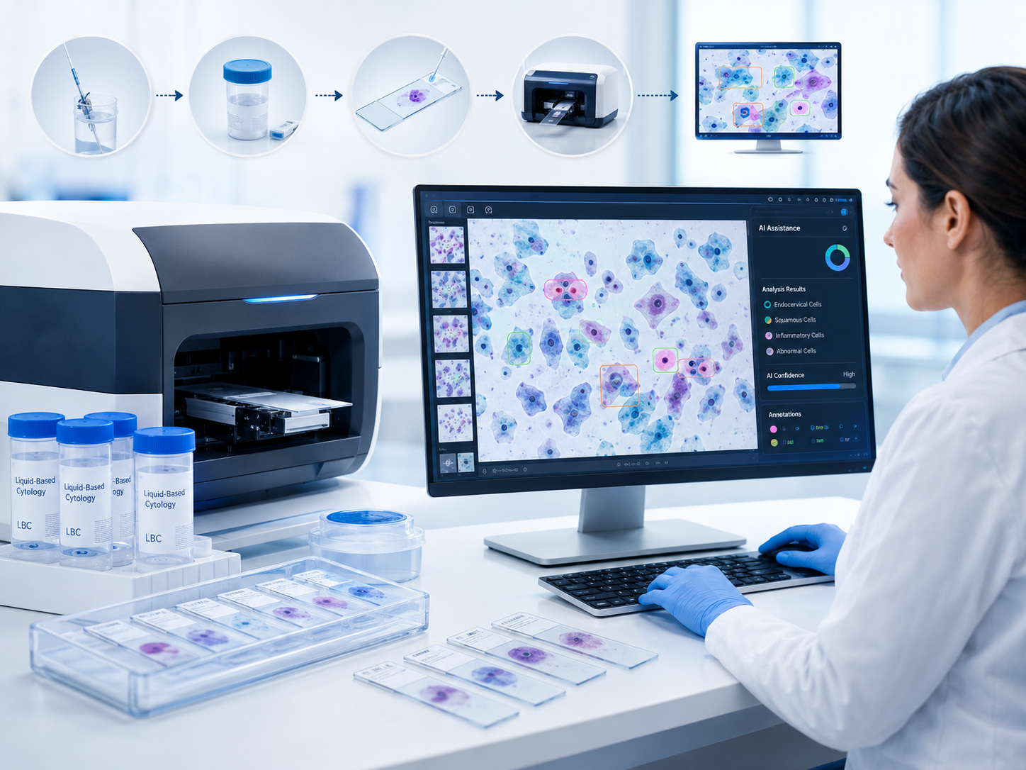
The cervical cytology screening workflow includes sample collection, slide preparation, staining, slide scanning or microscope review, interpretation, reporting, and clinical follow-up.
The workflow begins with cervical sample collection. The collected cells are then preserved and prepared for laboratory examination. In liquid-based cytology, the sample is suspended in a preservation solution, which helps create a cleaner and more uniform slide for review.
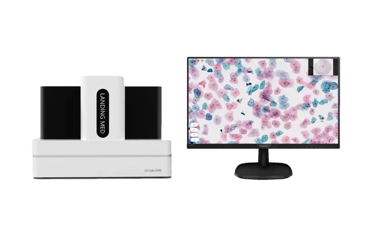
After preparation and staining, the slide is examined manually under a microscope or digitized using a cytology slide scanner. In a digital workflow, LandingMed’s LD Cyto2200 High Throughput Digital Pathology Slide Scanner can support high-throughput cervical cytology scanning with 200-slide capacity, 20X/40X magnification, automatic slide loading, scanning, and label identification.
A complete cervical cytology workflow should also include quality control. Laboratories need to manage slide labeling, case matching, image quality, reviewer workload, report consistency, and data traceability. These details are especially important for screening centers and reference laboratories that process large daily slide volumes.
Cervical cytology slide review is the process of examining stained cervical cells to identify normal findings, atypical changes, precancerous lesions, or suspicious malignant cells.
In traditional workflows, cytotechnologists and pathologists review slides under a microscope. They evaluate cellular features such as nuclear size, chromatin pattern, cytoplasmic changes, cell arrangement, and background quality. In digital workflows, the slide is converted into a whole slide image, allowing reviewers to zoom, navigate, annotate, and share cases on a workstation.
Reporting should be clear and standardized. A report may include sample adequacy, general interpretation, abnormal findings, and recommendations for follow-up based on local clinical guidelines. Digital cytology can improve traceability because scanned slides can be archived, re-opened, reviewed remotely, and used for training or quality assessment.
| Workflow Factor | Traditional Cervical Cytology | Digital Cervical Cytology |
|---|---|---|
| Slide review | Microscope-based manual review | Computer-based whole slide image review |
| Case sharing | Requires physical slide transfer | Easier remote consultation and case discussion |
| Traceability | More dependent on manual records | Supports image archive and annotations |
| Workload management | Mainly depends on human screening | Can support digital triage and workflow prioritization |
| AI compatibility | Limited without digitization | Can connect with AI-assisted analysis |
| Best fit | Small-volume or confirmatory review | Screening centers, hospitals, and high-volume labs |
AI assistance in cervical cytology uses algorithms to analyze digital slide images and help identify areas of interest for professional review.
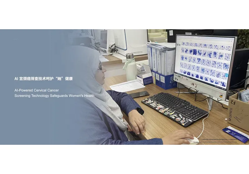
AI does not replace cytotechnologists or pathologists. Instead, it can support screening efficiency by highlighting suspicious cells, prioritizing cases, and reducing repetitive visual workload. This can be especially useful in large-scale cervical cancer screening programs where laboratories need to review many slides consistently.
Landing Med’s On-premise AI Cervical Cytology Solution uses scanned whole slide images from digital pathology scanners and supports AI-assisted analysis for cervical cytology workflows. The system is designed to help laboratories integrate sample preparation, slide scanning, AI analysis, pathologist review, and report issuance into a more complete digital workflow.
For laboratories planning AI-assisted cytology, implementation should be based on real workflow needs. Important factors include sample volume, scanner compatibility, data security, validation requirements, reviewer training, reporting process, and local regulatory compliance. AI should be used as a decision-support tool within a validated laboratory workflow, not as a substitute for professional judgment.
Cervical cytology remains a core method for cervical cancer screening because it helps identify abnormal cell changes before cancer develops. As screening demand grows, laboratories need workflows that are accurate, traceable, scalable, and easier to standardize.
Digital slide scanning and AI assistance can help laboratories improve cervical cytology workflow efficiency while supporting professional review. With liquid-based cytology consumables, LD Cyto2200 digital slide scanning, and AI-assisted cervical cytology solutions, LandingMed provides an integrated path for laboratories upgrading from manual screening to digital cervical cytology. To discuss a suitable workflow for your laboratory, contact Landing Med.
Cervical cytology is used to screen cervical cells for abnormal changes that may indicate precancerous lesions, cervical cancer risk, or the need for further testing.
A Pap smear is a common method of cervical cytology. Cervical cytology can include conventional Pap smears and liquid-based cytology workflows.
Sample adequacy affects whether enough cervical cells are available for reliable interpretation. Poor sample quality may lead to repeat testing or uncertain results.
Cervical cytology examines cell changes. HPV testing detects high-risk HPV infection. Many screening programs use cytology and HPV testing together depending on local guidelines.
Digital slide scanning converts glass slides into whole slide images, making review, archiving, remote consultation, training, and AI-assisted analysis easier.
No. AI can support screening and highlight areas of interest, but final interpretation should remain within a validated professional review workflow.


 fr
fr  es
es  CN
CN