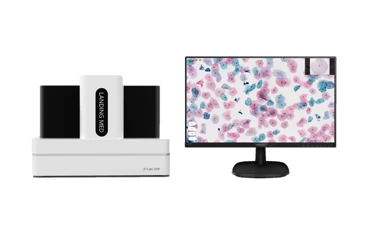
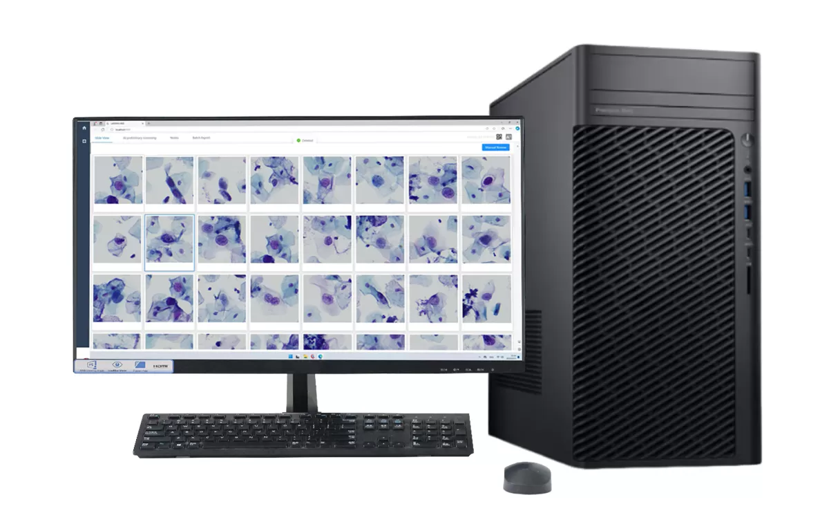
A cytology slide scanner is a digital pathology device that converts glass cytology slides into high-resolution whole slide images for computer-based review, storage, sharing, and AI-assisted analysis. It is widely used in cervical cytology, liquid-based cytology, screening centers, hospital pathology departments, and reference laboratories.
The need for efficient cytology screening is growing. According to the World Health Organization, cervical cancer caused about 660,000 new cases and 350,000 deaths worldwide in 2022. Digital cytology helps laboratories improve slide traceability, review efficiency, and collaboration while supporting large-scale screening programs.
For high-throughput digital cytology, LandingMed offers the LD Cyto2200 High Throughput Digital Pathology Slide Scanner, which supports 200-slide capacity, 20X/40X magnification, automatic slide loading, scanning, and label identification.
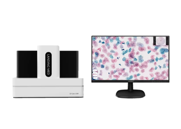
A cytology slide scanner is an automated imaging system that captures cells on glass slides and creates digital whole slide images for computer-based review.
Unlike histology slides, which usually contain tissue sections, cytology slides contain scattered cells from samples such as cervical specimens, body fluids, or fine-needle aspiration materials. Because cells may be distributed unevenly, cytology scanning requires stable focusing, clear cellular detail, and reliable image stitching.
In practice, a cytology slide scanner reduces repetitive manual microscope navigation. Once scanned, digital slides can be viewed, zoomed, annotated, archived, shared for consultation, and connected with AI-assisted analysis tools. The College of American Pathologists whole slide imaging validation guideline also highlights the importance of validating whole slide imaging systems before clinical diagnostic use.
Definition: A cytology slide scanner works by loading slides automatically, identifying the scan area, capturing high-resolution image tiles, stitching them into a whole slide image, and making the image available for digital review.
The workflow usually starts with batch slide loading. The scanner moves slides into the imaging position, reads or identifies slide labels, and links each image with the correct case information. This helps reduce manual matching errors.
Next, the system locates the cell area and performs focusing. Since cytology samples can vary in thickness and cell density, focus quality is critical. The scanner then captures image tiles at a selected magnification, commonly 20X or 40X, and software combines them into a complete digital slide.
After scanning, cytotechnologists and pathologists can review the slide on a workstation. In AI-assisted workflows, algorithms may help highlight suspicious areas or prioritize cases, but final interpretation still depends on professional review, validation, and local regulatory requirements.
Laboratories should compare slide capacity, magnification, automation, image quality, label recognition, software compatibility, AI integration, validation requirements, and service support before choosing a cytology slide scanner.
For high-volume labs, slide capacity is especially important. A compact scanner may fit a small laboratory, while a screening center may need batch scanning and automation. LandingMed LD Cyto2200 supports 200 slides, making it suitable for laboratories with larger daily slide volumes.
Magnification also affects workflow. 20X scanning supports efficient routine review, while 40X scanning provides more cellular detail. Automation features such as slide loading, scanning, and label identification can reduce manual workload and improve workflow consistency.
Labs planning digital transformation can also compare Landing Med’s digital pathology slide scanners and evaluate whether the scanner can integrate with AI-assisted cytology solutions. For regulatory planning, the U.S. FDA announcement on whole slide imaging for digital pathology shows that whole slide imaging has become an important part of digital pathology development.
| Factor | Cytology Slide Scanner | Traditional Microscope |
|---|---|---|
| Workflow | Automated scanning and digital review | Manual slide navigation |
| Capacity | Supports batch scanning; LD Cyto2200 supports 200 slides | Usually one slide at a time |
| Magnification | Digital imaging at 20X/40X | Depends on objective lenses |
| Sharing | Easy for remote consultation | Requires physical slide transfer |
| Traceability | Supports image archive and annotations | More dependent on manual records |
| AI integration | Can connect with AI-assisted workflows | Limited without digital imaging |
| Best fit | Screening centers and high-volume labs | Low-volume or confirmatory review |
A cytology slide scanner helps laboratories move from microscope-only review to digital cytology, whole slide imaging, and AI-assisted screening. It improves slide traceability, supports remote consultation, and helps high-volume laboratories build a more scalable review workflow.
For laboratories that need high-throughput cytology digitization, Landing Med LD Cyto2200 offers 200-slide capacity, 20X/40X magnification, automated slide loading, scanning, and label identification. To evaluate whether this configuration fits your laboratory workflow, contact Landing Med for digital pathology scanner options.
A cytology slide scanner is used to digitize glass cytology slides for digital review, storage, sharing, and AI-assisted analysis.
Yes. Cytology scanners focus on scattered cells, while histology scanners focus on tissue sections. Cytology scanning often requires stronger focus control and cellular detail.
20X helps with efficient overview and routine review, while 40X provides higher detail for cellular morphology.
No. AI can support screening efficiency, but professional review and validated workflows remain essential.
Cervical screening centers, hospital pathology departments, reference labs, and laboratories with large slide volumes can benefit from high-throughput scanning.
A lab should compare slide volume, magnification, scanning quality, automation, AI compatibility, software workflow, service support, and compliance requirements.


 fr
fr  es
es  CN
CN